

6.2%
RESILIA tissue*
Engineered to potentially allow the valve to last longer
With increasing life expectancy, long-lasting tissue valves are needed to limit the need for re-intervention in patients.1,2 The MITRIS RESILIA valve is built upon the trusted Carpentier-Edwards PERIMOUNT valve platform, which has over 20 years of published clinical durability.3–9 It incorporates bovine pericardial tissue that is less prone to tearing compared with porcine tissue, due to its higher collagen density.10–12
A retrospective study evaluating the durability of the Edwards mitral valves prior to the MITRIS RESILIA valve (comprised of 79% Carpentier-Edwards PERIMOUNT Magna Mitral Ease valve and the remainder on previous generations) found the cumulative incidence of mitral valve (MV) reinterventions performed for structural valve deterioration (SVD) was: †13
9.0%
at 12 years
When comparing amongst age groups, there was no significant difference in SVD amongst patients aged 40–70 years (P=0.1). 13
The MITRIS RESILIA valve takes this foundation a step further with RESILIA tissue, designed to offer enhanced anti-calcification technology that will potentially allow the valve to last even longer. RESILIA tissue is bovine pericardial tissue with a special integrity preservation technology that effectively eliminates free aldehydes while protecting and preserving the tissue.
Porcine and pericardial valves have different modes of failure14
Porcine valves are prone to acute failure due to cusp tears, which causes sudden valvular insufficiency. This can result in acute heart failure and haemodynamic instability, increasing the surgical urgency.4,14,15 In re-do valve replacement surgeries, patients with prior porcine valves have significantly higher rates of urgent re-intervention compared to those with pericardial valves.15
In contrast, pericardial valves tend to fail from stenosis through a slower process of progressive calcification.4,14,15 The difference in failure speed likely accounts for the higher rate of surgical urgency observed in porcine valves vs pericardial valves:15

Adapted from Tang GHL, et al. 2007.15
Addressing calcification – the primary mechanism of pericardial valve failure4,14,15 – has driven the innovation behind RESILIA tissue.
The MITRIS RESILIA valve incorporates Edwards Lifesciences’ latest generation bovine pericardial tissue
RESILIA tissue has a special integrity preservation technology that effectively eliminates free aldehydes, a key factor in tissue calcification, potentially allowing the valve to last longer.
The anti-calcification properties of the Carpentier-Edwards PERIMOUNT valve with RESILIA tissue were compared to the PERIMOUNT valve without RESILIA tissue in the mitral position of juvenile sheep, and RESILIA tissue demonstrated favourable anti-calcification properties. §16
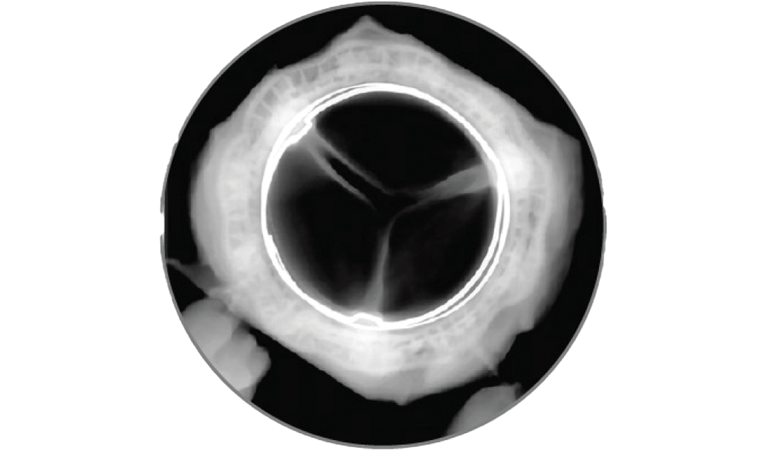
RESILIA tissue
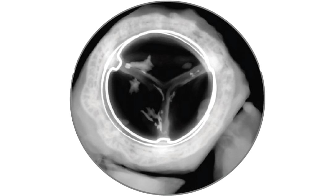
Control tissue
Adapted from Flameng W, et al. 2015.16
Anti-calcification technology

RESILIA tissue is supported by a strong and growing body of clinical evidence in the aortic position of up to 7 years,21,22 and now the latest data supports its use in the mitral position23
COMMENCE mitral 5-year clinical trial (median age 70 years): 23
98.7%
freedom from SVD
(95% CI: 96.1, 100%)
97.1%
freedom from reoperation
(95% CI: 93.1, 100%)
Find out how the MITRIS RESILIA valve could impact your patients’ lifetime management of mitral valve disease
*Clinical data on surgical aortic valves with RESILIA tissue up to 7-year follow-up have been published, with additional follow-up to 10 years in progress.21
†A retrospective study at Northwestern and Michigan Medicine assessed the durability of pericardial bioprosthetic mitral valves by age and reintervention risk. 1,544 patients underwent mitral valve replacement with PERIMOUNT/PERIMOUNT Theon (n=225), Magna Mitral (n=97), and Magna Mitral Ease (n=1,220). Patients had a mean age of 66±13 years; 59% were female, 57.4% NYHA III/IV, 57% had heart failure, and 48% had atrial fibrillation. Durability was defined as freedom from reintervention (surgical or valve-in-valve).13
‡743 patients underwent redo valve surgery (mitral, aortic or both). During re-operation, 814 valves were explanted, of which 34% were mechanical (n=274), 48% were porcine (n=390) and 18% were pericardial (n=150).15
§45 female juvenile sheep (<6 months of age) weighing between 22–38 kg underwent mitral valve replacement. Sheep were randomised to either receive the 25 mm Carpentier-Edwards PERIMOUNT mitral valve (6900P) (n=23) or the PERIMOUNT mitral valve (6900P) incorporating RESILIA tissue (n=22). At week 1 and 8 months post-implant, sheep were imaged with transthoracic echocardiography, after which valves were excised and examined radiographically, histologically and chemically. 16
Abbreviations
AOA, alpha-amino oleic acid; CI, confidence interval; MV, mitral valve; NYHA, New York Heart Association; SVD, structural valve deterioration.
References
- Wise J. BMJ. 2024;385:q1126.
- Bourguignon T, et al. Eur J Cardiothorac Surg. 2018;54(2):302–9.
- Aupart MR, et al. J Thorac Cardiovasc Surg. 1997;113(3):492–8.
- Jamieson WR, et al. J Thorac Cardiovasc Surg. 1999;118(2):297–304.
- Murakami T, et al. J Heart Valve Dis. 1996;5(1):45–9.
- Poirer NC, et al. Ann Thorac Surg. 1998;66(6 Suppl):S57–61.
- Neville PH, et al. Ann Thorac Surg. 1998;66(6 Suppl):S143–7.
- Marchand MA, et al. Ann Thorac Surg. 2001;71(5 Suppl):S236–9.
- Bourguignon T, et al. J Thorac Cardiovasc Surg. 2014;148(5):2004–11.
- Bezuidenhout D, et al. Biotechnol Appl Biochem. 2009;54(3):133–40.
- Uchino G, et al. Eur J Cardiothorac Surg. 2022;62(1):ezab506.
- Liao K, et al. Artif Organs. 1992;16(4):361–5.
- Romano M, et al. J Thorac Cardiovasc Surg. 2024;168(5):1448–58.
- Keshishi M, et al. Cardiovasc Pathol. 2023;65:107516.
- Tang GHL, et al. Ann Thorac Surg. 2007;83(3):938–45.
- Flameng W, et al. J Thorac Cardiovasc Surg. 2015;149:340–5.
- Vyavahare N, et al. Circulation. 1997;95(2):479–88.
- Chen W, et al. J Biomed Mater Res. 1994;28(12):1485–95.
- Tod TJ, et al. J Mater Sci Mater Med. 2016;27(1):8.
- Dove J, et al. Carpentier-Edwards ThermaFix Process: a method for extracting calcium binding sites from pericardial tissue. Edwards Lifesciences LLC. 2006.
- Beaver T, et al. J Thorac Cardiovasc Surg. 2024;168(3):781–91.
- Bartus K, et al. Eur J Cardiothorac Surg. 2021;59:434–41.
- Heimansohn DA, et al. JTCVS Open. 2023;15:151–163.
Medical device for professional use.
Medical device for professional use.
For a listing of indications, contraindications, precautions, warnings, and potential adverse events, please refer to the Instructions for Use (consult eifu.edwards.com where applicable).
PP--EU-11857 v1.0